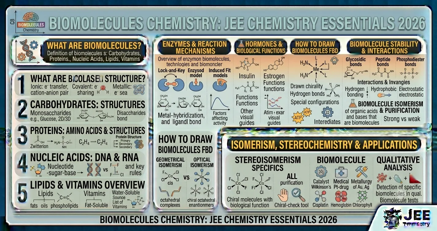
Chemistry · JEE Main & Advanced 2026 · Biomolecules

Biomolecules notes, structures & PYQs for JEE 2026

Complete chapter — carbohydrates (glucose, fructose structures, mutarotation), amino acids & proteins (zwitterions, peptide bond, protein structures), enzymes, vitamins (classification & deficiency), nucleic acids (DNA, RNA, nucleotides, double helix), 150+ worked examples, and JEE PYQs with solutions.

SECTIONS
8
SOLVED EXAMPLES
150+
READ TIME
~4h
COST
₹0

Table of Contents

Jump to any section
Biomolecules JEE Notes, Carbohydrates, Proteins, DNA, PYQs
Biomolecules — JEE Notes, Structures, PYQs
1

Introduction & Classification of Carbohydrates

Biomolecules are the complex organic molecules that form the basis of life. They include carbohydrates, proteins, lipids, nucleic acids, vitamins, and hormones. This chapter holds significant weightage in JEE, particularly for direct structural questions and matching lists (Vitamins/Amino acids).

1.1 What are Carbohydrates?

Carbohydrates are polyhydroxy aldehydes or ketones, or substances that yield such compounds on hydrolysis. They are primarily produced by plants during photosynthesis.

General Formula

\[ C_n(H_2O)_n \quad \text{or} \quad C_x(H_2O)_y \]

Exceptions: Rhamnose (C₆H₁₂O₅) and Deoxyribose (C₅H₁₀O₄) do not follow the general formula but are still carbohydrates.

1.2 Classification of Carbohydrates

TypeHydrolysis ProductExamples
MonosaccharidesCannot be hydrolyzed furtherGlucose, Fructose, Ribose
Oligosaccharides (Di/Tri/Tetra)Yield 2-10 monosaccharidesSucrose, Maltose, Lactose, Raffinose
PolysaccharidesYield >10 monosaccharidesStarch, Cellulose, Glycogen

Important — Reducing vs Non-Reducing Sugars

  • Reducing Sugars: Have a free aldehyde (–CHO) or ketone (>C=O) group. Reduce Tollens' and Fehling's reagent. Examples: All monosaccharides (glucose, fructose), Disaccharides like Maltose and Lactose.
  • Non-Reducing Sugars: Lack free anomeric carbon (both anomeric carbons are involved in glycosidic bond). Examples: Sucrose (table sugar).
2

Monosaccharides: Glucose & Fructose

Monosaccharides are the simplest carbohydrates. Glucose (an aldohexose) and Fructose (a ketohexose) are the most important from a JEE perspective.

2.1 Structure of D-Glucose

Open Chain Structure (Fischer Projection)

Glucose has the molecular formula C₆H₁₂O₆. Its open chain shows an aldehyde group at C1 and hydroxyl groups on C2-C5.

\[ \text{CHO} - (\text{CHOH})_4 - \text{CH}_2\text{OH} \]

Chiral Carbons: C₂, C₃, C₄, C₅ (Total = 4). Therefore, glucose has $2^4 = 16$ stereoisomers (8 D and 8 L forms).

Most Repeated PYQ — Cyclic Structure of Glucose (Haworth & Pyranose)

The open-chain structure of glucose does not explain some of its properties (like lack of Schiffs test and existence of α and β forms). The cyclic structure is formed when the -OH at C₅ attacks the aldehyde group at C₁, forming a six-membered ring called Pyranose.

  • α-D-Glucose: The -OH at the anomeric carbon (C₁) is on the opposite side of the -CH₂OH group (trans).
  • β-D-Glucose: The -OH at the anomeric carbon (C₁) is on the same side as the -CH₂OH group (cis).
  • Anomeric Carbon: The carbon that was previously a carbonyl carbon (C₁ in aldoses, C₂ in ketoses) and becomes a new stereocenter in the cyclic form.

Most Important — Mutarotation

When pure α-D-glucose (specific rotation +112°) is dissolved in water, its specific rotation changes to +52.5°. Similarly, pure β-D-glucose (+19°) also changes to +52.5° in water. This spontaneous change in optical rotation is called Mutarotation.

\[ \alpha\text{-D-Glucose} \rightleftharpoons \text{Open Chain} \rightleftharpoons \beta\text{-D-Glucose} \] \[ (+112^\circ) \quad\quad\quad\quad\quad\quad (+19^\circ) \] \[ \text{Equilibrium mixture} = +52.5^\circ \]

2.2 Structure of D-Fructose

Fructose is a ketohexose (C₆H₁₂O₆). It has a ketone group at C₂.

\[ \text{CH}_2\text{OH} - \text{CO} - (\text{CHOH})_3 - \text{CH}_2\text{OH} \]

Cyclic Form: The -OH at C₅ attacks the C₂ ketone to form a five-membered ring called Furanose.

Chiral Carbons: C₃, C₄, C₅ (Total = 3). Fructose has $2^3 = 8$ stereoisomers.

PYQ — JEE Main 2024 (Jan 31 Shift 1)

Question: The number of chiral carbon atoms present in the open-chain structure of glucose and fructose respectively are:

(a) 4 and 3

(b) 4 and 4

(c) 5 and 4

(d) 3 and 4

Solution:

Glucose: CHO-(CHOH)₄-CH₂OH

- C₁ is CHO (sp², not chiral)

- C₂, C₃, C₄, C₅ are CHOH (each attached to 4 different groups) → 4 chiral carbons

- C₆ is CH₂OH (not chiral)

Fructose: CH₂OH-CO-(CHOH)₃-CH₂OH

- C₁ is CH₂OH (not chiral)

- C₂ is C=O (sp², not chiral)

- C₃, C₄, C₅ are CHOH → 3 chiral carbons

- C₆ is CH₂OH (not chiral)

\[\text{Glucose: 4, Fructose: 3}\]

Answer: (a) 4 and 3

3

Disaccharides & Polysaccharides

Disaccharides yield two monosaccharides on hydrolysis, linked by a glycosidic bond (an ether linkage formed by the loss of water between two anomeric carbons or one anomeric and one alcoholic -OH).

3.1 Glycosidic Linkage in Disaccharides

Most Repeated PYQ — Sucrose vs Maltose vs Lactose Linkage

DisaccharideMonosaccharidesGlycosidic BondReducing Nature
Sucrose (Cane Sugar)Glucose + Fructoseα-1,2-glycosidic (C1 of glucose & C2 of fructose)Non-Reducing (no free anomeric carbon)
Maltose (Malt Sugar)Glucose + Glucoseα-1,4-glycosidic (C1 of one & C4 of other)Reducing (one free anomeric C present)
Lactose (Milk Sugar)Glucose + Galactoseβ-1,4-glycosidic (C1 of galactose & C4 of glucose)Reducing (one free anomeric C present)

Key Insight: For a disaccharide to be reducing, one of the anomeric carbons must be free (not involved in the glycosidic bond) so it can open to form an aldehyde/ketone.

3.2 Polysaccharides

Polysaccharides are long chains of monosaccharides. They act as energy reserves (Starch, Glycogen) or structural components (Cellulose).

Starch

Polymer of α-D-Glucose.

  • Amylose: Linear, α-1,4 linkage (20%).
  • Amylopectin: Branched, α-1,4 & α-1,6 linkage (80%).
  • Food reserve in plants.

Cellulose

Polymer of β-D-Glucose.

  • Straight chain, β-1,4 linkage.
  • Major structural component of plant cell walls.
  • Humans lack cellulase enzyme, cannot digest it.

Glycogen

Polymer of α-D-Glucose.

  • Highly branched (more than amylopectin).
  • α-1,4 & α-1,6 linkage.
  • Animal starch (stored in liver & muscles).

PYQ — JEE Main 2023

Question: Which of the following statements is incorrect about sucrose?

(a) It is a non-reducing sugar.

(b) On hydrolysis, it gives glucose and fructose.

(c) The glycosidic linkage is between C1 of glucose and C4 of fructose.

(d) It does not exhibit mutarotation.

Solution:

Sucrose Structure: The glycosidic linkage in sucrose is formed between the anomeric carbon of glucose (C1) and the anomeric carbon of fructose (C2). This is an α-1,2-glycosidic linkage.

Statement (c) claims the linkage is C1 of glucose and C4 of fructose, which is incorrect. (C4 of fructose is not the anomeric carbon; C2 is).

Because both anomeric carbons are tied up in the bond, there is no free aldehyde/ketone group, making it non-reducing (a) and preventing mutarotation (d). Hydrolysis yields glucose + fructose (b), known as invert sugar.

\[\text{Linkage is C1 (glucose) - C2 (fructose)}\]

Answer: (c) The glycosidic linkage is between C1 of glucose and C4 of fructose.

4

Amino Acids & Zwitterion

Proteins are the building blocks of life and are polymers of α-amino acids. There are 20 standard amino acids, out of which 10 are essential (must be taken through diet) and 10 are non-essential (synthesized by the body).

4.1 Structure of Amino Acids

Amino acids contain an amino group (–NH₂) and a carboxyl group (–COOH). In α-amino acids, the amino group is attached to the α-carbon (the carbon adjacent to the –COOH group).

\[ \overset{+}{H_3}N - CH_2 - COO^- \quad \text{(Glycine - simplest)} \] \[ \text{General: } H_2N - \overset{\alpha}{C}H(R) - COOH \]

Except for glycine (where R = H), all other amino acids are chiral (except Proline which is a secondary amine).

Most Repeated PYQ — Zwitterion & Isoelectric Point

In aqueous solution or crystalline state, the –COOH group loses a proton and the –NH₂ group gains a proton, forming a dipolar ion called Zwitterion.

\[ H_2N - CH(R) - COOH \rightleftharpoons \overset{+}{H_3}N - CH(R) - COO^- \]
  • Isoelectric Point (pI): The pH at which the amino acid exists as a neutral zwitterion (no net charge). At this point, it has minimum solubility and does not migrate in an electric field.
  • In Acidic Medium (pH < pI): Exists as a cation ($^+H_3N–CH(R)–COOH$) and migrates to the cathode.
  • In Basic Medium (pH > pI): Exists as an anion ($H_2N–CH(R)–COO^-$) and migrates to the anode.

PYQ — JEE Main 2022

Question: Amino acids exist as zwitterions. At the isoelectric point, the amino acid:

(a) migrates towards the cathode

(b) migrates towards the anode

(c) does not migrate in an electric field

(d) has maximum solubility

Solution:

At the isoelectric point, the concentration of the positive form equals the concentration of the negative form.

The structure is perfectly balanced as a zwitterion: $^+H_3N–CH(R)–COO^-$

Because the net charge is zero, the amino acid experiences no net electrostatic force and does not migrate towards either electrode in an electric field.

Also, due to balanced charges and maximum internal salt formation, its solubility in water is minimum (not maximum).

\[\text{Net charge} = 0 \implies \text{No migration}\]

Answer: (c) does not migrate in an electric field

5

Proteins & Peptide Bond

Proteins are polypeptides. When the –COOH group of one amino acid reacts with the –NH₂ group of another, a peptide bond is formed with the elimination of water.

5.1 Peptide Bond Formation

\[ H_2N - CH(R_1) - COOH + H_2N - CH(R_2) - COOH \rightarrow H_2N - CH(R_1) - \overset{\displaystyle O}{\overset{\displaystyle ||}{C}} - NH - CH(R_2) - COOH + H_2O \]

The linkage –CO–NH– is called a peptide bond. It is an amide linkage. Dipeptides have 2 amino acids, tripeptides have 3, and so on. Proteins typically have >50 amino acids.

5.2 Structure of Proteins

High PYQ Frequency — Levels of Protein Structure

1. Primary Structure

The specific linear sequence of amino acids linked by peptide bonds. The sequence determines the biological function. Insulin was the first protein to have its primary structure determined (by Sanger).

2. Secondary Structure

The spatial arrangement of the polypeptide chain, stabilized by hydrogen bonds between backbone amide and carbonyl groups. Two main types:

  • α-Helix: Right-handed coiled structure, stabilized by intrachain H-bonds (e.g., Keratin in hair).
  • β-Pleated Sheet: Extended polypeptide chains lying side by side, stabilized by interchain H-bonds (e.g., Silk fibroin).

3. Tertiary Structure

The overall 3D shape of a single polypeptide chain, folded compactly. Stabilized by weak interactions (H-bonds, Van der Waals, ionic bonds) and strong disulfide (–S–S–) linkages.

4. Quaternary Structure

The arrangement of two or more polypeptide chains (subunits) into a multi-subunit protein. E.g., Hemoglobin has 4 subunits (2 α and 2 β).

5.3 Denaturation of Proteins

Denaturation is the loss of the secondary, tertiary, and quaternary structures of a protein without breaking the peptide bonds (primary structure remains intact).

Agents causing denaturation: Heat, changes in pH, urea, heavy metal salts (Hg²⁺, Pb²⁺), organic solvents.

Example: Coagulation of egg white on boiling (loss of globular structure of albumin).

PYQ — JEE Main 2021

Question: The secondary structure of a protein refers to:

(a) Sequence of α-amino acids

(b) Hydrogen bonding between peptide bonds leading to α-helix or β-pleated sheet

(c) Bonding between R groups of amino acids

(d) Complete 3D arrangement of the polypeptide chain

Solution:

(a) refers to Primary structure.

(b) Hydrogen bonding between the backbone -NH and -C=O groups of the peptide linkages leads to localized folding into α-helix or β-pleated sheets. This defines Secondary structure.

(c) Bonding between R groups (e.g., disulfide bonds, ionic bonds) stabilizes the Tertiary structure.

(d) The complete 3D arrangement is also Tertiary structure.

\[\text{Secondary structure} \implies \text{H-bonds between backbone}\]

Answer: (b) Hydrogen bonding between peptide bonds leading to α-helix or β-pleated sheet

6

Enzymes & Vitamins

6.1 Enzymes

Enzymes are biological catalysts. Almost all enzymes are globular proteins (exception: Ribozymes are RNA). They catalyze biological reactions with high specificity and efficiency.

Mechanism of Enzyme Action

An enzyme (E) binds to a substrate (S) to form an enzyme-substrate complex (ES), which then converts to the product (P) and releases the enzyme.

\[ E + S \rightleftharpoons ES \rightarrow E + P \]
  • Active Site: The specific region of the enzyme where the substrate binds.
  • Lock and Key Model (Fischer): The active site is rigid and fits exactly one substrate.
  • Induced Fit Model (Koshland): The active site is flexible and molds itself to fit the substrate.

6.2 Vitamins

Vitamins are organic compounds required in small quantities for normal metabolic functions. They are not synthesized by the body (except Vitamin D) and must be obtained from the diet.

Most Repeated PYQ — Classification of Vitamins

VitaminNameSolubilityDeficiency Disease
Vitamin ARetinolFatNight blindness, Xerophthalmia
Vitamin B1ThiamineWaterBeri-Beri
Vitamin B2RiboflavinWaterCracked lips, Dermatitis
Vitamin B6PyridoxineWaterConvulsions, Anemia
Vitamin B12CyanocobalaminWaterPernicious anemia
Vitamin CAscorbic acidWaterScurvy (bleeding gums)
Vitamin DCalciferolFatRickets (children), Osteomalacia (adults)
Vitamin ETocopherolFatInfertility, Muscle weakness
Vitamin KPhylloquinoneFatDelayed blood clotting

JEE Shortcut — Remembering Fat-Soluble Vitamins

Mnemonic: "A DEK" (Vitamins A, D, E, K are Fat-soluble).

All B-complex vitamins and Vitamin C are water-soluble. Because they are water-soluble, they cannot be stored in the body and are excreted in urine. Fat-soluble vitamins can be stored in the liver and adipose tissue.

PYQ — JEE Main 2024 (Apr 4 Shift 2)

Question: Which of the following vitamins is a fat-soluble vitamin and its deficiency causes night blindness?

(a) Vitamin B1

(b) Vitamin C

(c) Vitamin A

(d) Vitamin B12

Solution:

1. Identify the deficiency disease: Night blindness is caused by a deficiency of Vitamin A (Retinol). (It is essential for the formation of rhodopsin, the visual pigment).

2. Check solubility: The fat-soluble vitamins are A, D, E, and K. Vitamin A is indeed fat-soluble.

B1 and B12 are water-soluble (B-complex) and cause Beri-Beri and Pernicious anemia respectively. Vitamin C is water-soluble and causes Scurvy.

\[\text{Vitamin A (Retinol) - Fat-soluble}\]

Answer: (c) Vitamin A

7

Nucleic Acids (DNA & RNA)

Nucleic acids are the genetic material of cells. There are two types: Deoxyribonucleic Acid (DNA) and Ribonucleic Acid (RNA). They are polymers of nucleotides.

7.1 Nucleoside vs Nucleotide

Most Repeated PYQ — Nucleotide Composition

  • Nucleoside = Nitrogenous Base + Pentose Sugar
  • Nucleotide = Nitrogenous Base + Pentose Sugar + Phosphate Group

Nucleotides are the monomeric units of nucleic acids, linked by phosphodiester bonds (between the 3'-OH of one sugar and the 5'-phosphate of the next).

7.2 Components of Nucleic Acids

1. Nitrogenous Bases

  • Purines (2 rings): Adenine (A), Guanine (G)
  • Pyrimidines (1 ring): Cytosine (C), Thymine (T) - only in DNA, Uracil (U) - only in RNA.

2. Pentose Sugar

  • β-D-2-Deoxyribose in DNA
  • β-D-Ribose in RNA
  • Difference: DNA sugar lacks an -OH group at the 2' position (has only -H).

3. Phosphate Group

Linked to the 5'-carbon of the sugar via an ester bond. Provides the acidic nature to nucleic acids.

7.3 DNA Double Helix (Watson-Crick Model)

Most Repeated PYQ — Base Pairing & H-Bonds

DNA consists of two polynucleotide chains running antiparallel (one 3'→5', the other 5'→3'), coiled into a right-handed double helix.

Chargaff's Rule (Base Pairing):

  • Adenine (A) always pairs with Thymine (T) via 2 Hydrogen bonds (A = T).
  • Guanine (G) always pairs with Cytosine (C) via 3 Hydrogen bonds (G ≡ C).
  • A = T and G = C (amount of purines = amount of pyrimidines).
  • A + G = T + C (Total purines = Total pyrimidines).

Stability: The G-C pair is more stable than the A-T pair because it has one extra hydrogen bond. Higher G-C content means higher melting point for DNA.

PYQ — JEE Main 2024 (Jan 29 Shift 2)

Question: In a DNA segment, the amount of Adenine is 28%. What will be the percentage of Guanine in the same segment?

(a) 28%

(b) 22%

(c) 56%

(d) 44%

Solution:

According to Chargaff's rule in a double-stranded DNA:

Amount of Adenine (A) = Amount of Thymine (T)

Amount of Guanine (G) = Amount of Cytosine (C)

Also, A + G = T + C = 50%

Given: A = 28%

Therefore, T = 28% (since A = T)

Total A + T = 28% + 28% = 56%

The remaining percentage is for G + C = 100% - 56% = 44%

Since G = C, G = 44% / 2 = 22%

\[ \%G = \frac{100 - 2(A)}{2} = \frac{100 - 56}{2} = 22\% \]

Answer: (b) 22%

Critical JEE Trap — DNA vs RNA

  • DNA contains Thymine; RNA contains Uracil instead.
  • DNA sugar is 2-Deoxyribose; RNA sugar is Ribose.
  • DNA is usually double-stranded; RNA is usually single-stranded.
  • DNA is present in the nucleus (and mitochondria); RNA is formed in the nucleus but functions in the cytoplasm.
8

Hormones & Biomolecules Summary

8.1 Hormones

Hormones are biochemical messengers produced by endocrine glands. They are released directly into the bloodstream and act on specific target organs.

Classification of Hormones

  • Steroid Hormones: Derived from cholesterol. Examples: Testosterone, Estradiol, Progesterone, Cortisol. They are lipid-soluble and can pass through cell membranes.
  • Peptide/Protein Hormones: Made of amino acids. Examples: Insulin (regulates blood glucose), Glucagon, Oxytocin, Vasopressin.
  • Amino Acid Derivatives: Examples: Epinephrine (Adrenaline), Norepinephrine, Thyroxine.

8.2 Tests for Carbohydrates & Proteins

Test NameReagentObservationIndicates
Molisch's Testα-Naphthol + Conc. H₂SO₄Violet ring at junctionGeneral carbohydrate
Tollens' TestAmmoniacal AgNO₃Silver mirrorReducing sugar
Fehling's TestFehling's A & BRed ppt (Cu₂O)Reducing sugar
Barfoed's TestCu(OAc)₂ in AcOHRed pptMonosaccharides only
Ninhydrin TestNinhydrin reagentBlue/Purple colorProteins & α-amino acids
Biuret TestNaOH + CuSO₄Violet colorProteins (Peptide bonds)
Xanthoproteic TestConc. HNO₃Yellow colorAromatic amino acids

PYQ — JEE Main 2023

Question: Insulin and Glucagon belong to which class of hormones?

(a) Steroid hormones

(b) Amino acid derivatives

(c) Peptide hormones

(d) Iodoamino acids

Solution:

1. Insulin is a peptide hormone produced by the β-cells of the pancreas. It lowers blood glucose levels by promoting glycogen synthesis.

2. Glucagon is also a peptide hormone produced by the α-cells of the pancreas. It increases blood glucose levels by breaking down glycogen.

Both are made of amino acid chains (polypeptides). Steroid hormones include cortisol and testosterone. Thyroxine is an iodoamino acid. Epinephrine is an amino acid derivative.

\[\text{Insulin & Glucagon = Peptide Hormones}\]

Answer: (c) Peptide hormones

Previous Year Questions Analysis

JEE Main (Last 10 Years)

  • ✓ DNA/RNA Base Pairing & H-bonds: 22%
  • ✓ Vitamin Classification & Deficiency: 18%
  • ✓ Glucose/Fructose Structure (Chiral Carbons): 15%
  • ✓ Amino Acids & Zwitterion/Isoelectric Point: 12%
  • ✓ Glycosidic Linkage (Sucrose/Maltose): 10%
  • ✓ Protein Structures (α-helix): 8%
  • ✓ Nucleotide vs Nucleoside: 7%
  • ✓ Hormones & Enzymes: 8%

JEE Advanced (Last 10 Years)

  • ✓ Matching Vitamins with Structure/Deficiency: 25%
  • ✓ Cyclic Structure of Carbohydrates: 20%
  • ✓ Identifying Anomeric Carbon/Mutarotation: 15%
  • ✓ Amino Acid Reactions (Peptide bond count): 15%
  • ✓ DNA double helix stability (G-C vs A-T): 15%
  • ✓ Tests for Biomolecules: 10%

Top 10 Most Repeated Question Types

  1. Calculate percentage of nitrogenous bases in DNA given one base % (Chargaff's rule).
  2. Match vitamins with their chemical names and deficiency diseases.
  3. Count the number of chiral carbons in glucose, fructose, or nucleotides.
  4. Identify the isoelectric point behavior of amino acids.
  5. Differentiate between α-D-glucose and β-D-glucose (Haworth projections).
  6. Identify reducing/non-reducing sugars and the nature of their glycosidic linkages.
  7. Differentiate between nucleoside and nucleotide components.
  8. Number of hydrogen bonds in DNA base pairing.
  9. Protein secondary structure stabilization (Intrachain H-bonds in α-helix).
  10. Classify hormones into steroid vs peptide categories.

Weightage Analysis

JEE Main: 8-12 marks (2-3 questions)
JEE Advanced: 4-8 marks (1-2 questions)
Difficulty Level: Easy to Moderate
Time Required: 3-4 hours practice

Practice Problem Set

Level 1: Basic (JEE Main Standard)

  1. How many chiral carbons are present in β-D-(-)-Fructofuranose?
  2. Which vitamin deficiency causes Pernicious anemia?
  3. What is the linkage type in cellulose?
  4. An amino acid has an isoelectric point of 6.0. Will it migrate to the cathode or anode at pH 8.0?
  5. In a DNA molecule, if Adenine constitutes 30%, what is the percentage of Guanine?

Level 2: Intermediate (JEE Main/Advanced)

  1. A tetrapeptide contains 4 amino acids. How many peptide bonds and water molecules are involved in its formation?
  2. Identify the non-reducing sugar among: Glucose, Maltose, Lactose, Sucrose. Explain why.
  3. Which structural level of protein is disrupted during denaturation? Does the primary structure remain intact?
  4. What is the structural difference between the sugars present in DNA and RNA?
  5. Match the enzymes: Trypsin, Amylase, Pepsin, Lipase with their respective substrates.

Level 3: Advanced (JEE Advanced/Olympiad)

  1. A DNA segment has 1000 base pairs. If it has 30% G-C content, calculate the total number of H-bonds in the segment.
  2. Draw the Haworth structure of α-D-glucopyranose and identify the anomeric carbon.
  3. Why is sucrose called an invert sugar? Explain the optical rotation changes during its hydrolysis.
  4. An amino acid exists as a cation in a solution. If the pH is adjusted to exactly 7.0, it stops migrating. What is its isoelectric point, and what form does it take?
  5. Explain why starch gives a blue-black color with iodine, while cellulose does not.

> FAQ

Frequently Asked Questions

What is mutarotation in carbohydrates?

Mutarotation is the change in specific optical rotation of a freshly prepared solution of a pure crystalline carbohydrate. It occurs because the alpha and beta anomers of the sugar interconvert through the open-chain aldehyde form in solution, reaching an equilibrium mixture.

What is a zwitterion?

A zwitterion is a dipolar ion containing both a positive (ammonium, -NH3+) and a negative (carboxylate, -COO-) charge within the same molecule. Amino acids exist as zwitterions at their isoelectric point, where they have no net electrical charge and minimum solubility.

What is the difference between nucleoside and nucleotide?

A nucleoside consists of a nitrogenous base (purine or pyrimidine) attached to a pentose sugar (ribose or deoxyribose). A nucleotide is a nucleoside with an additional phosphate group attached to the 5'-carbon of the sugar. Nucleotides are the building blocks of nucleic acids (DNA and RNA).

Which vitamin is fat-soluble?

Fat-soluble vitamins are Vitamins A, D, E, and K. They are absorbed along with fats in the diet and can be stored in the body's fatty tissues. Water-soluble vitamins (B-complex and C) cannot be stored and must be regularly consumed.

How many hydrogen bonds are present between Guanine and Cytosine in DNA?

There are three (3) hydrogen bonds between Guanine (G) and Cytosine (C) in a DNA double helix. Adenine (A) and Thymine (T) are connected by two (2) hydrogen bonds. This makes G-C pairs stronger and more stable than A-T pairs.

What is the secondary structure of proteins?

The secondary structure of proteins refers to the spatial arrangement of the polypeptide chain, stabilized by hydrogen bonds between the backbone amide (-NH) and carbonyl (-C=O) groups. The most common types are the alpha-helix (right-handed coil) and beta-pleated sheet.

Related Chemistry Notes

Complete Biomolecules Mastery Guide for JEE 2026

Why This Chapter is Important

Biomolecules is one of the most scoring chapters in JEE Organic Chemistry:

  • 2-3 questions in almost every JEE Main session
  • Highly factual — requires less conceptual derivation and more memorization
  • Matching list questions in JEE Advanced frequently come from Vitamins/Amino acids
  • Direct structural questions on glucose, DNA base pairing, and zwitterions
  • Application-based questions on enzyme mechanisms and protein denaturation

Topic-wise Weightage Analysis

Carbohydrates (Glucose, Sucrose) 35%
Nucleic Acids (DNA/RNA) 25%
Vitamins 20%
Amino Acids & Proteins 20%

Must-Remember Formulas & Facts

Carbohydrates

Glucose: Aldohexose (4 chiral C)

Fructose: Ketohexose (3 chiral C)

Sucrose: α-1,2 linkage (Non-reducing)

Maltose: α-1,4 linkage (Reducing)

Lactose: β-1,4 linkage (Reducing)

Cellulose: β-1,4 (Plant structure)

Starch/Glycogen: α-1,4 & α-1,6

Amino Acids

General: H₂N–CH(R)–COOH

Zwitterion: ⁺H₃N–CH(R)–COO⁻

Peptide bond: –CO–NH–

Essential amino acids: 10

Nucleic Acids

Nucleoside = Base + Sugar

Nucleotide = Base + Sugar + Phosphate

A = T (2 H-bonds)

G ≡ C (3 H-bonds)

DNA: Deoxyribose, Thymine

RNA: Ribose, Uracil

Vitamins (Fat-Soluble)

A - Retinol (Night blindness)

D - Calciferol (Rickets)

E - Tocopherol (Infertility)

K - Phylloquinone (Blood clotting)

Biomolecules — Complete JEE Study Guide

Biomolecules is a highly scoring chapter in JEE Organic Chemistry, covering the structure and function of carbohydrates, proteins, amino acids, enzymes, vitamins, and nucleic acids. While largely factual, this chapter demands precise memorization of structural details (like the Haworth structure of glucose, the difference between α and β anomers, and the glycosidic linkages in disaccharides) to avoid losing marks in tricky questions.

The most important topics for JEE from this chapter are mutarotation (the interconversion of α-D-glucose and β-D-glucose through the open-chain form), zwitterion and isoelectric point of amino acids, Chargaff's base pairing rule in DNA (A=T with 2 hydrogen bonds, G≡C with 3 hydrogen bonds), and the classification of vitamins into fat-soluble (A, D, E, K) and water-soluble (B, C) with their respective deficiency diseases.

Students should focus on mastering the distinction between nucleosides (Base + Sugar) and nucleotides (Base + Sugar + Phosphate), the difference between DNA (Deoxyribose, Thymine) and RNA (Ribose, Uracil), and the four levels of protein structure (primary, secondary, tertiary, quaternary). The secondary structure, particularly the α-helix (right-handed coil stabilized by intrachain hydrogen bonds) and β-pleated sheet (interchain hydrogen bonds), is a frequent exam favorite.

For JEE Advanced, expect matching list questions connecting vitamins to their chemical names (e.g., Retinol, Thiamine, Calciferol, Ascorbic acid) and deficiency diseases. Additionally, knowing the number of chiral carbons in open-chain and cyclic forms of glucose (4) and fructose (3), as well as identifying non-reducing sugars (sucrose) based on the glycosidic linkage involving both anomeric carbons, is crucial for securing full marks in this chapter.